- Home
- News & Events
- News
AI, Advanced Microscopy and Faster Disease Diagnosis: Yaraslau Padrez Becomes a PhD
On 17 September, Yaraslau Padrez, a researcher at the FTMC Department of Molecular Compound Physics, celebrated his birthday with a remarkable gift to himself – on the very same day he defended his PhD.
His thesis topic is “Machine Learning-based Diagnosis of Cancer and Fibrosis with Second Harmonic Generation Microscopy” (academic supervisor: Dr Renata Karpicz; academic consultant: Dr Danielis Rutkauskas).
Congratulations to our colleague – wishing him every success to move forward!
The 2024 Nobel Prize in Physics was awarded to two US scientists for their development of machine learning (ML), which laid the foundation for artificial intelligence (AI). FTMC is also at the forefront – and Yaraslau’s work, contributing to more accurate and faster diagnosis of serious diseases, is proof of that.
For example, in cancer treatment, increasing attention is being paid not only to new therapeutic methods but also to early diagnosis. Advances in technology and the application of AI in medicine open up new possibilities – computerized analysis of tissue images can become a reliable assistant to pathologists and provide additional diagnostic insights.
In his research, Padrez focuses on two diseases – thyroid cancer and pulmonary fibrosis. How does it work? Simply put, the necessary tissues were taken from the lungs of laboratory rats and from the thyroid glands of human patients, then illuminated with an infrared laser to determine their properties and the disease status. This method is called second harmonic generation (SHG) microscopy.
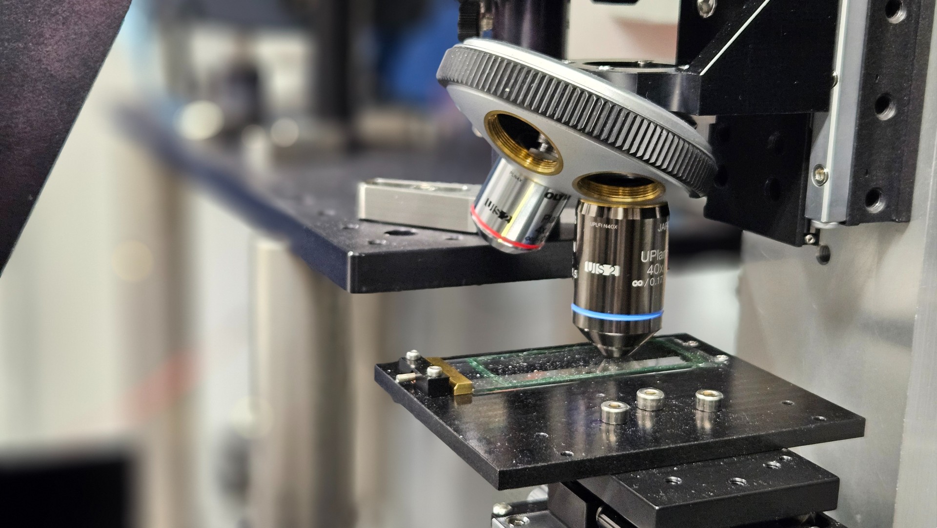
(SHG microscope used for sample imaging. Photo: FTMC)
The FTMC researcher combines this microscopy with AI models, and one of the key achievements is that papillary and follicular carcinomas (different types of thyroid cancer) were recognized with 84.7% accuracy.
“The dissertation is about applying ML techniques on wide-field SHG images sets to automate and to improve the diagnosis of diseases by analyzing collagen fibers in tissue samples,” says Yaraslau.
Collagen protein plays a crucial role here. When a tumour begins to form in the thyroid gland, the body responds: it covers the unwanted mass with a collagen capsule that isolates the tumour from the surrounding tissues. If doctors see that this capsule remains intact, the tumour is considered benign. But if the capsule is altered or disrupted, the tumour is malignant. It is therefore vital to detect these changes in time.
Collagen also affects the lungs: when it starts accumulating in the walls of blood vessels, these walls thicken, lose elasticity, stiffen – and breathing becomes difficult.
SHG microscopy enables researchers to effectively observe collagen in thyroid capsules and lung tissues – and to obtain valuable data from its imaging.
.jpg)
(Malignant thyroid tumour with collagen capsule around it. Photo: FTMC)
“We successfully identified and quantitatively described specific stages of pulmonary arterial hypertension (PAH) by analyzing collagen changes in rat lung tissue. We showed that so-called unsupervised ML (like PCA and k-means) can uncover a hidden textural heterogeneity in the collagen capsule of thyroid tumours.
I think my work could be interesting to the wider public because it sits at the cutting edge of using AI to make healthcare more precise, objective, and efficient, with direct benefits for patients with cancer and chronic lung disease,” explains Dr Padrez.
The dissertation can be accessed via this link.
FTMC Information